
Fungal Skin Infections: What They Look Like and How to Treat Them
When people search for fungal skin infections pictures, they’re usually trying to confirm what they’re seeing on their own body. Skin fungus pictures from medical references show a wide variety of appearances — rings, patches, scaling, blistering — and knowing what distinguishes one type from another determines which treatment actually works. Fungal skin infection pictures consistently show specific patterns based on the organism and the body area affected, which makes visual recognition a practical first step.
Skin fungus images in medical literature cover several distinct infections: ringworm, athlete’s foot, jock itch, nail fungus, and candida (yeast) infections. Each is a fungal infection of skin, but they affect different areas, look different, and respond to different antifungal approaches. This guide gives you a clear picture of each condition.
Ringworm (Tinea Corporis)
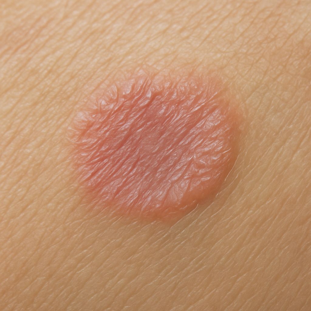
Despite its name, ringworm has nothing to do with worms. It’s a fungal skin infection producing a characteristic ring-shaped rash with a raised, scaly, reddish border and clearer skin in the center. Fungal skin infection pictures of ringworm show this distinctive ring clearly, though in early stages it may appear as just a red, circular patch. It typically itches and spreads outward over days to weeks.
Ringworm spreads by direct contact with infected people, animals, or contaminated surfaces. Treatment is OTC antifungal cream (clotrimazole, miconazole, or terbinafine) applied twice daily for 2–4 weeks. Do not stop treatment when visible improvement appears — stopping early allows regrowth.
Athlete’s Foot (Tinea Pedis)
Athlete’s foot is among the most common fungal infections of skin. Skin fungus pictures of this condition show peeling, redness, and scaling between the toes and on the sole of the foot. The moccasin type creates thick, scaly skin across the entire sole. The vesicular type produces blisters. All forms itch and can have a mildly unpleasant odor from secondary bacterial involvement.
Treatment: keep feet dry, wear moisture-wicking socks, treat with an OTC antifungal for at least 4 weeks. Chronic or widespread cases may need oral terbinafine.
Jock Itch (Tinea Cruris)
Jock itch affects the groin and inner thighs, with a reddish-brown rash that has a raised, defined border. Fungal skin infection pictures of tinea cruris show the classic horseshoe or semicircular pattern around the upper thigh. It’s more common in men and in those who sweat heavily. OTC antifungal creams work effectively. Keep the area dry and avoid tight-fitting clothing during treatment.
Candida (Yeast) Skin Infections
Candida infections produce a different appearance than tinea infections. Skin fungus images of candidiasis show bright red, sometimes weeping patches in skin folds: under the breasts, in the groin, armpits, or between fingers in people who work with their hands in water frequently. There are often satellite lesions — small red spots surrounding the main patch. Candida thrives in warm, moist environments.
Treatment involves keeping the area dry and applying antifungal cream (clotrimazole or nystatin). Severe or recurring candida infections may indicate an underlying condition like diabetes or immune suppression — a doctor visit is warranted.
Nail Fungus (Onychomycosis)
Nail fungus is technically a fungal infection of skin — the nail bed and surrounding tissue. It produces yellowing, thickening, and crumbling of the nail, usually starting at the tip and moving toward the base. Skin fungus pictures of onychomycosis show discoloration ranging from pale yellow to brown-black in severe cases. OTC treatments rarely penetrate the nail sufficiently. Prescription oral terbinafine or itraconazole for 6–12 weeks is the standard treatment for complete clearance.
When to See a Doctor
OTC antifungal treatment works for most mild to moderate infections. See a doctor when: symptoms don’t improve after 2–4 weeks of treatment, the infection is spreading rapidly, you have diabetes or immune suppression, or the affected area shows signs of secondary bacterial infection (increased warmth, purulent discharge, spreading red streaks).
Bottom line: Most fungal skin infections are recognizable from skin fungus pictures and respond well to OTC antifungals when treated consistently and for the full recommended duration. Match the treatment to the specific fungal infection type — tinea infections need a different antifungal than candida in some cases. When in doubt or when treatment fails, a dermatologist can confirm the organism with a simple scraping and culture.


Leave a Reply