
Precancerous skin lesions are abnormal growths that have not yet become malignant but carry a measurable risk of progressing to skin cancer without treatment. Precancerous skin conditions span several specific types, the most common being actinic keratosis, which results from years of cumulative UV exposure. Understanding precancerous skin cancer risk means recognizing that removal is both preventive and curative at this stage. Pre cancer skin lesions are the body’s warning signal, and catching them at this point is the optimal outcome of regular dermatological monitoring. Skin precancer is treatable in most cases with simple in-office procedures, and the range of treatment options available means there is usually a suitable approach for each patient’s needs and skin type.
This guide explains the types of precancerous lesions, how they are diagnosed, and what treatment is most appropriate for each presentation.
What Are Precancerous Skin Lesions?
Precancerous skin lesions are areas of abnormal cell growth that have not yet invaded deeper skin layers or spread but show cellular changes consistent with early carcinogenesis. They arise from cumulative DNA damage, most commonly from ultraviolet radiation, though some types involve HPV infection or chronic inflammation. Without treatment, a percentage of precancerous skin lesions progress to squamous cell carcinoma (SCC) or, less commonly, basal cell carcinoma (BCC) or melanoma. Estimates vary, but actinic keratosis, the most common type, converts to SCC at a rate of around 0.1-10% per lesion per year, with higher risk in immunocompromised patients.
What Is Actinic Keratosis and What Does It Look Like?
Actinic keratosis (AK) appears as rough, scaly patches ranging from pink to red to brown, typically found on sun-exposed areas: the face, scalp (particularly in bald areas), ears, backs of the hands, and forearms. The texture is the most characteristic feature; they feel like sandpaper and are easier to feel than to see in early stages. Some AKs develop a prominent scale or “cutaneous horn” of hardened keratin. They may itch, burn, or bleed with minor trauma. Multiple AKs on sun-damaged skin indicate field cancerization, where the surrounding skin tissue has accumulated sufficient UV damage that new lesions will continue to form without broader treatment.
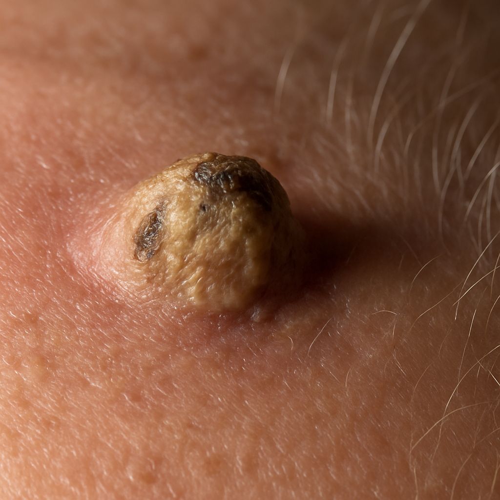
What Other Types of Pre Cancer Skin Lesions Exist?
Bowen’s disease (squamous cell carcinoma in situ) appears as a persistent, slowly enlarging red plaque with scaling. Unlike AK, Bowen’s disease involves the full thickness of the epidermis and is considered the most advanced form of pre cancer skin lesion before true invasion occurs. Dysplastic nevi (atypical moles) are precancerous skin lesions in the melanocytic category; those with severe atypia on biopsy carry a meaningful risk of progressing to melanoma and require excision. Leukoplakia, a white patch in the mouth or on the lip, is a mucosal precancerous condition related to HPV and tobacco use.
Who Is at Highest Risk of Developing Skin Precancer?
The strongest risk factors for precancerous skin cancer are fair skin with a history of sunburns, cumulative sun exposure over decades, use of tanning beds, and living in high-altitude or high-UV-index environments. Immunosuppression from organ transplant medication, HIV, or long-term corticosteroid use significantly increases both the rate of AK development and the risk of conversion to invasive cancer. A family history of non-melanoma skin cancer and male sex are also established risk factors, as men are less likely to use sunscreen consistently and may have more occupational UV exposure.
How Are Precancerous Skin Conditions Treated?
Cryotherapy with liquid nitrogen is the most common treatment for individual AKs. The dermatologist applies a short burst of liquid nitrogen that destroys the abnormal cells with freezing; a small blister forms and heals within one to two weeks. Topical therapies including 5-fluorouracil (5-FU) cream, imiquimod, and ingenol mebutate treat areas of field cancerization by destroying multiple precancerous cells across a sun-damaged area simultaneously. Photodynamic therapy (PDT) uses a photosensitizing agent applied to the skin and then activated by a specific wavelength of light; it is effective for widespread AKs and provides cosmetically superior results on the face. Curettage (scraping) with or without electrodesiccation is used for thicker, hyperkeratotic lesions.
How Do You Monitor Skin for Precancerous Changes?
Annual full-body skin examinations by a dermatologist remain the gold standard for early detection. Between appointments, monthly self-examination of the face, scalp, neck, hands, forearms, and other sun-exposed areas helps catch changes between professional checks. Any new rough, scaly, or non-healing patch that persists for more than four weeks deserves a dermatologist appointment. People with a personal history of AK or non-melanoma skin cancer benefit from more frequent monitoring, typically every six months. Daily broad-spectrum SPF 30+ sunscreen use and sun-protective clothing are the most effective preventive interventions for people at elevated risk.
Bottom line: Precancerous skin lesions are manageable when identified and treated early. Actinic keratosis is the most common form of skin precancer, and multiple treatment options can eliminate it before it progresses. Regular dermatologist monitoring combined with daily sun protection is the most reliable strategy for preventing the development of new pre cancer skin lesions.


Leave a Reply